Picture yourself touching a hot stove or stepping on a sharp pebble. In a fraction of a second, sensory receptors in your skin fire signals racing up your arm or leg, warning your brain to pull away before you even realize what happened.
We tend to think of our nervous system as a simple network of electrical wires carrying signals effortlessly from one place to another. But neurons are much more complex and dramatic than standard copper wires. Why doesn’t a tiny, gentle brush against your skin trigger a massive pain response? Why must incoming electrical signals accumulate before a neuron finally decides to fire?
We take our lightning-fast reflexes for granted, assuming our nerves react instantly to everything. Take the interactive neuron simulation below to test how electrical pulses build up membrane potential, and discover the exact moment an action potential is born.
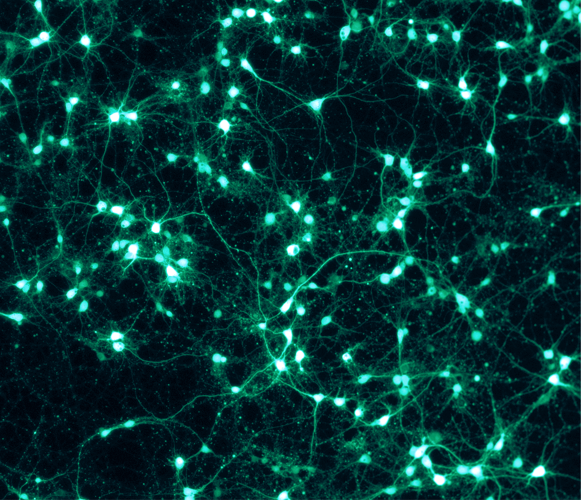
Try it
Do you predict that sending a single weak pulse will cause the neuron to fire, or will you need to send multiple quick pulses to push the membrane potential across the threshold?
Make a neuron fire
membrane potential, mVAfter playing with the pulse buttons, your results reveal the fundamental rule of neural communication: the all-or-none law. When you send a single weak pulse, the membrane potential nudges upward briefly before fading back to its resting state of negative seventy millivolts because the signal failed to reach the threshold.
However, when you send four quick pulses in rapid succession, temporal summation kicks in. The electrical charges stack on top of each other until the voltage at the axon hillock crosses the critical threshold. Suddenly, voltage-gated sodium channels fly open, a massive spike shoots up, and an action potential fires down the axon. If you don’t reach the threshold, nothing happens at all; once you cross it, the neuron fires at full force.
The science
The resting state and ion channels
To understand how a neuron fires, we have to look at what happens at rest. A resting neuron maintains a negative electrical charge inside its cell membrane, sitting around -70 mV. This resting potential is maintained by sodium-potassium pumps that actively shuttle positive sodium ions out of the cell and potassium ions in, creating a charged battery waiting to be discharged.
The axon hillock and threshold
When dendrites receive chemical signals from neighboring cells, they open ligand-gated ion channels, letting small amounts of positive ions flow inward. These graded potentials travel across the cell body toward the axon hillock, which acts as the neural decision center. If the cumulative voltage reaching the axon hillock hits the threshold (around -55 mV), an action potential is triggered.
The anatomy of a neuron
Orchestrating this electrochemical cascade requires intense coordination across specialized cell regions, which you can explore on the interactive diagram on this page:
- Dendrites (Inputs): Branching structures that receive chemical neurotransmitters from other neurons.
- Cell Body (Soma): Houses the nucleus and metabolic machinery that keep the neuron alive.
- Axon Hillock: The decision gateway where incoming graded potentials are summed up and the threshold for firing is evaluated.
- Axon (Wrapped in Myelin): A long cable that transmits the electrical action potential rapidly away from the cell body, insulated by fatty myelin sheaths that help signals jump efficiently between nodes of Ranvier.
- Terminals (Outputs): The end branches that release neurotransmitters across the synapse to communicate with the next cell.
Notice in the Neuroscientifically Challenged breakdown (2-Minute Neuroscience: Action Potential) how sodium and potassium ions rush across the cell membrane in a precise, choreographed sequence during depolarization and repolarization. Action potentials are not just static sparks; they are dynamic chemical waves moving down the axon.
Why it matters
While simulating neuron spikes on a computer screen feels like a clean physics puzzle, the exact same cellular mechanisms power every thought, movement, emotion, and sensation you experience. Disruptions in ion channels and action potential conduction underlie major neurological disorders, including multiple sclerosis, epilepsy, and peripheral neuropathies.
Furthermore, research in neuroscience and public health demonstrates that cellular communication and neural plasticity are heavily influenced by environmental factors. Chronic sleep deprivation, high stress, and neurotoxins can impair synaptic efficiency and neural signaling. Understanding the science of how neurons fire helps shift our perspective away from viewing the brain as a static organ, highlighting instead how dynamic and sensitive our nervous system is to our daily environment and well-being.
Try this at home
You don’t need a neurobiology laboratory to explore reflex arcs and neural signaling. Try a classic reaction time test with a friend: have them hold a standard ruler vertically just above your open hand, and let it drop unexpectedly. Catch it as fast as you can and measure the distance. That quick grab relies on your eyes sending action potentials to your brain, which then sends signals through motor neurons down your spinal cord and arm to your hand muscles. Discuss with a classmate how fast biological wires can move.
Sources
- Hodgkin, A. L., & Huxley, A. F. (1952). A quantitative description of membrane current and its application to conduction and excitation in nerve. Journal of Physiology, 117(4), 500–544.
- 2-Minute Neuroscience: Action Potential by Neuroscientifically Challenged. YouTube.
- Purves, D. et al. (2018). Neuroscience (6th ed.). Sinauer Associates.